
Intravascular Imaging
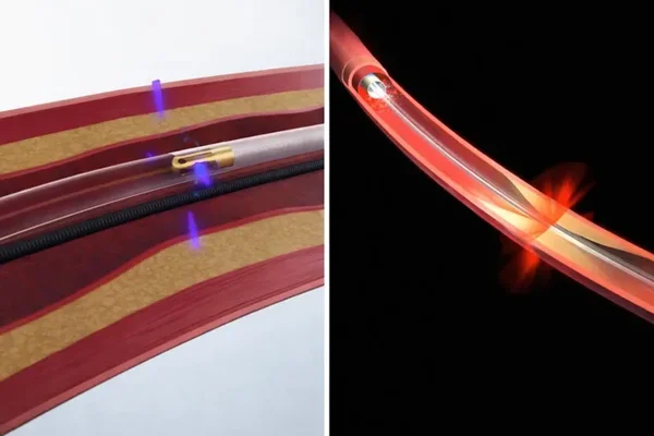
Intravascular Imaging
Accurate assessment of coronary artery disease is essential for selecting the right treatment and achieving optimal results during interventional cardiac procedures. Intravascular imaging using IVUS and OCT provides detailed images from inside the coronary arteries, helping cardiologists understand the size, structure, and severity of arterial blockages.
Dr. Kamlesh Raut is an experienced Interventional Cardiologist in Dwarka who provides comprehensive evaluation and advanced cardiac care. Depending on the patient's condition and clinical requirements, intravascular imaging may be used to support treatment planning and guide coronary interventions.
What Is Intravascular Imaging?
Intravascular imaging is an advanced technique that allows the cardiologist to examine the inside of a blood vessel using a specialized imaging catheter. Unlike conventional angiography, which provides a two-dimensional outline of the artery, intravascular imaging can provide more detailed information about the arterial wall, plaque, and vessel dimensions.
Two commonly used technologies are:
- IVUS – Intravascular Ultrasound
- OCT – Optical Coherence Tomography
Both techniques can provide valuable information during the evaluation and treatment of coronary artery disease.
What Is IVUS?
Intravascular Ultrasound (IVUS) uses high-frequency sound waves to create images of the inside of the coronary arteries. A small ultrasound transducer is positioned on a catheter and carefully advanced into the coronary artery.
IVUS can help assess:
- Vessel diameter and dimensions
- Plaque buildup
- Severity and characteristics of coronary lesions
- Extent of arterial narrowing
- Stent position and expansion
- Vessel wall characteristics
This information can assist the cardiologist in selecting an appropriate treatment strategy and optimizing coronary stent placement when required.
What Is OCT?
Optical Coherence Tomography (OCT) is an intravascular imaging technique that uses near-infrared light to produce high-resolution images of the coronary arteries.
OCT provides detailed visualization of the artery and can help assess features such as plaque characteristics, the inner vessel lining, and stent positioning. Its high resolution can be particularly useful for evaluating the result of coronary interventions.
IVUS vs OCT
Both IVUS and OCT are valuable intravascular imaging technologies, but they have different characteristics.
IVUS uses ultrasound and can provide information about vessel dimensions and plaque burden. It can also be useful when deeper vessel structures need to be assessed.
OCT provides very high-resolution images and can offer detailed visualization of the inner layers of the artery and coronary stents.
The choice between IVUS and OCT depends on the patient's coronary anatomy, clinical situation, procedure being performed, and the information required by the interventional cardiologist.
When Is IVUS or OCT Recommended?
Intravascular imaging may be considered in selected patients undergoing evaluation or treatment for coronary artery disease. It can be particularly useful when:
- Coronary artery anatomy is complex
- The severity of a blockage is difficult to determine with angiography alone
- Accurate vessel sizing is required
- Stent placement needs detailed assessment
- Stent expansion or positioning needs to be evaluated
- There is uncertainty regarding the characteristics of a coronary lesion
- Additional information is needed to guide an interventional procedure
Not every patient undergoing coronary angiography or angioplasty requires IVUS or OCT. The cardiologist determines whether intravascular imaging is appropriate based on the individual case.
How Is Intravascular Imaging Performed?
IVUS or OCT is generally performed as part of a catheter-based coronary procedure. After appropriate preparation and local anaesthesia, a catheter is introduced through a blood vessel and guided towards the coronary arteries.
A specialized imaging catheter is then positioned within the artery. For IVUS, ultrasound waves generate images of the vessel. For OCT, near-infrared light is used to create high-resolution images.
The images are reviewed by the interventional cardiologist to understand the coronary artery anatomy and guide treatment when required.
If the patient is undergoing angioplasty and stenting, intravascular imaging can also help evaluate the treated artery and assess the position and expansion of the stent.
Benefits of IVUS & OCT
Detailed Coronary Assessment
IVUS and OCT provide information that may not be fully visible through conventional angiography alone.
Better Treatment Planning
Detailed vessel measurements and plaque assessment can help the cardiologist select an appropriate treatment approach.
Stent Optimization
Intravascular imaging can help assess stent placement, expansion, and contact with the artery wall.
High-Resolution Imaging
OCT provides particularly detailed images of the inner structures of the coronary artery and stent.
Personalized Interventional Care
The information obtained through IVUS or OCT can help the cardiologist tailor an intervention according to the patient's coronary anatomy.
Why Choose Dr. Kamlesh Raut?
Dr. Kamlesh Raut is an experienced Interventional Cardiologist in Dwarka with advanced training in cardiac medicine and interventional cardiology. He completed his MD in Medicine from AIIMS, New Delhi, followed by DM in Cardiology from SGPGIMS, Lucknow.
He currently serves as Senior Consultant – Interventional Cardiologist at Max Super Speciality Hospital, Dwarka. His professional experience includes clinical cardiac care, interventional cardiology, academic work, and research.
Dr. Raut follows an evidence-based and patient-centred approach, carefully evaluating coronary artery disease before selecting an appropriate diagnostic or treatment strategy. When clinically indicated, advanced intravascular imaging such as IVUS or OCT can provide additional information to support complex coronary interventions.
Frequently Asked Questions
1. What does IVUS stand for?
IVUS stands for Intravascular Ultrasound. It uses ultrasound technology to create detailed images of the inside of coronary arteries.
2. What does OCT stand for?
OCT stands for Optical Coherence Tomography. It uses near-infrared light to create high-resolution images of the coronary arteries.
3. Is IVUS the same as coronary angiography?
No. Coronary angiography provides images of blood flow and the outline of the coronary arteries, while IVUS provides detailed imaging from inside the artery, including information about the vessel wall and plaque.
4. Why is IVUS or OCT used during angioplasty?
IVUS or OCT can provide detailed information about vessel size, plaque, and stent positioning. This may help the interventional cardiologist plan and optimize the procedure.
5. Is intravascular imaging required for every angioplasty?
No. IVUS and OCT are used selectively. The cardiologist decides whether intravascular imaging is beneficial based on the patient's condition, coronary anatomy, and complexity of the procedure.
6. Who provides IVUS and OCT-guided cardiac care in Dwarka?
Dr. Kamlesh Raut is an experienced Interventional Cardiologist in Dwarka who provides specialized evaluation and interventional cardiac care. The suitability of IVUS or OCT is determined based on individual clinical requirements.